


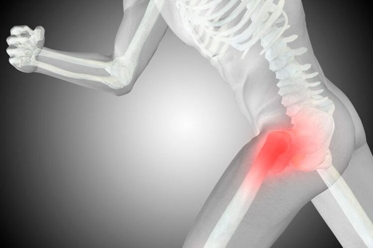
Coxarthrosis is a common degenerative-dystrophic disease of the hip in which, due to age-related changes or other factors, there is a gradual destruction of the articulation of the femoral head and pelvis. It is accompanied by pain and limited range of motion of varying severity, depending on the stage of development. And if in the early stages it is possible to cure the spasm by conservative methods, in stage 3 it is possible to save the situation and maintain the functioning of the hip joint, that is, to avoid disability, just byperform activities.
It belongs to the number of diseases of the joints and can be accompanied by the development of similar processes in other joints, and this pathology accounts for about 12% of all diseases of the musculoskeletal system. But the term "coxarthrosis" can only be used to describe degenerative-dystrophic changes in the hip joint.
What is coxarthrosis?
Coxarthrosis is a complex disease of one or both hip joints in which the cartilage covering the femoral head and joint capsule is destroyed, leading to a reduction in joint cavity size. As the disease progresses, the appearance of deformation of the surface of the bones and the formation of bony masses on them, called osteoblasts, is observed.
Coxarthrosis is the second most common disease of the musculoskeletal system. Usually, only gonarthrosis is diagnosed, i. e. a degenerative-dystrophic change in the knee joint. However, the probability of disability in coxarthrosis is significantly higher.
The entire hip joint is enclosed in a particular case, it is called a capsule. It has a so-called synovial membrane, which produces synovial fluid. This fluid is essential for the normal functioning of the joint, as it not only lubricates the hyaline cartilage, but also provides it with nutrients.
Normally, cartilage is constantly worn away and is restored immediately due to the continuous process of regeneration, which is carried out with the help of substances that enter it from the synovial fluid. But with injuries or age-related changes, the rate of regeneration decreases, which leads to a gradual wear of the hyaline cartilage and the development of coxarthrosis.
This is due to changes in the amount of synovial fluid produced and its composition. Under the influence of adverse factors, it becomes thicker and is produced in smaller volumes. As a result, the synovial fluid is no longer able to supply the hyaline cartilage with all the substances it needs in the proper amounts, leading to dehydration and rapid thinning. Gradually, the strength and elasticity of the cartilage decreases, areas of delamination of the fibers form it, cracks form in it, and the thickness also decreases. These changes can be noticed in instrumental diagnostic methods, in particular, the narrowing of the joint space draws attention to itself.
The narrowed joint space leads to increased friction between the bony structures that make up the hip joint and increased pressure on the already degraded hyaline cartilage. This causes more damage to it, affecting joint functioning and human condition, as deformed areas prevent the femoral head from sliding easily within the joint. As a result, there are symptoms of coxarthrosis.
If left untreated, the pathological changes worsen and the hyalinized cartilage wears down. Then, in some areas, it completely disappears, resulting in exposed bone and a sharp increase in the load on the joint. Because when moving inside the tibia, the femoral head will rub directly against the bone, which causes the appearance of severe pain and limited mobility. In this case, the pressure of the bony structures on each other leads to the formation of bony masses that develop on their surface.
The bone cells that have formed can have sharp parts that can damage the muscles and ligaments around the hip joint. This leads to the appearance of intense pain both directly in the joint and groin area, buttocks and thighs. As a result, the patient removes the injured leg, puts less stress on it, and tries to avoid making unnecessary movements with it. This causes the development of atrophy, which aggravates dyskinesia and eventually leads to limping.
Reason
There are many reasons for the development of coxarthrosis, although in rare cases it occurs against the background of the absence of any prerequisites. In this case, they talk about the presence of primary or idiopathic coxarthrosis. In the vast majority of cases, secondary coxarthrosis is diagnosed, which becomes a logical consequence of some disease or changes in the state of the musculoskeletal system. It can be provoked by:
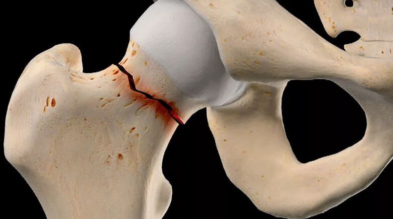
- injuries of the hip joint of various nature, including fracture, dislocation, bruise, sprain or tear of the ligament;
- hard physical labor, professional sports, especially weightlifting, skydiving, skydiving;
- sedentary lifestyle;
- overweight, which significantly increases the load on the hip joint;
- foci of chronic infection in the body;
- birth defects of the hip joint, such as dysplasia or dislocation;
- metabolic and endocrine disorders, in particular gout, diabetes, especially in the form of decompensation;
- aseptic necrosis of the femoral head, which may result from femoral neck fractures, especially when treated conservatively;
- inflammatory arthritis, including rheumatoid arthritis, bursitis, tendinitis;
- diseases of the spine;
- genetic predisposition;
- the presence of bad habits, especially smoking.
However, the main cause of coxarthrosis remains the inevitable age-related changes, and the presence of the above factors only increases the likelihood and rate of development of the disease.
Symptoms of coxarthrosis
The disease is characterized by a gradual progression with a systematic increase in the intensity of symptoms. Therefore, in the early stages, the disease may have no symptoms or only occasionally cause anxiety to the patient, but then the hip condition worsens, leading to an increase in the severity of the symptoms. of genital warts up to unbearable pain and significant limitation of mobility. .
So, degenerative-dystrophic changes in the hip are accompanied by:
- Pain of varying degrees, arises initially after exertion or walking and subsides after rest. Gradually, the severity of the pain syndrome increases, it occurs more often, lasts longer, and the interval between the moment when force is applied to the joint and the occurrence of pain is reduced. After that, the pain appeared almost continuously, even at rest, and became unbearable. Pain increases at any stage of disease development when hypothermia and heavy lifting are characteristic.
- Limited mobility of the hip, manifested initially by minor difficulties in performing rotational movements of the leg. Over time, morning stiffness appears and disappears after the patient "dissolves". This may be accompanied by the appearance of edema in the hip joint. As the disease progresses, mobility limitations become more pronounced and persistent, i. e. they do not go away after warm-up. The patient notices a decrease in the range of leg movements, and then a complete loss of the ability to perform some movements.
- Cracks in the hip joint that occur during walking or physical activity, especially during stretching. It becomes the result of friction of bare bone structures against each other, resulting in a sharp increase in pain.
- Thigh muscle spasm, resulting in radiating pain in the thigh. This may be the result of a combination of various intra-articular disorders, including nerve compression, overstretching of the ligaments surrounding the joint, as well as the development of bursitis, i. e. synovitis. is inflammation of the synovial membrane and accumulation of inflammatory substances. effusion in the cavity of the hip joint.
- Soreness, initially as a result of the patient's unconscious desire to reduce the load on the diseased joint and shift weight to the healthy leg to avoid the appearance or intensifying of pain, and later to the development of muscle contraction. The latter phenomenon occurred in the later stages of coxarthrosis and led to the fact that the patient could not fully straighten the leg and, moreover, keep it in this position. As a result, the lower extremities with the hip are constantly affected in a slightly curvilinear position, causing lameness.
- Decreased leg length, which occurs mainly with severe degenerative-dystrophic changes in the hip, is not only accompanied by narrowing of joint space, but also due to flattened femoral head and muscle atrophy. As a result, the affected leg becomes 1 cm or more shorter than the healthy leg.
Coxarthrosis can affect both one hip and both at the same time. But if in the first case the symptoms of the disease are observed only on one side, then in the second case they will be not only bilateral, but also different in intensity. It depends on the degree of destruction of each hip joint.
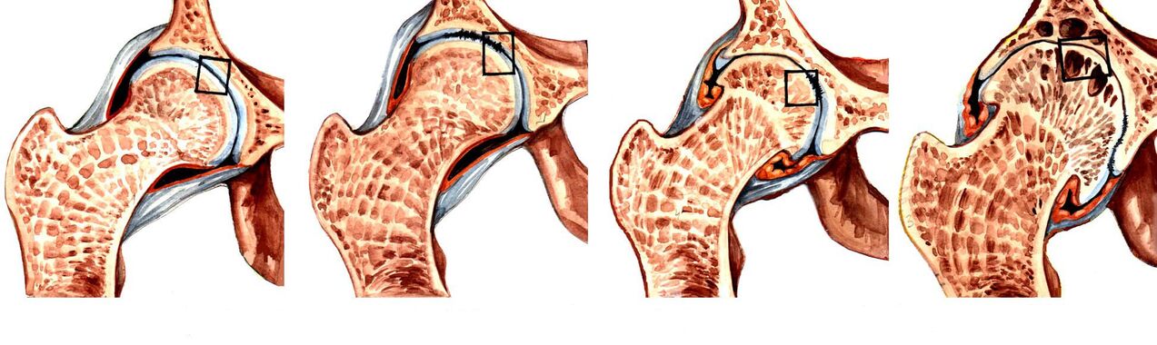
Coxarthrosis level
The nature of the manifestations of the disease depends on the stage of its development. There are 3 degrees of coxarthrosis in total, of which the first is considered the easiest. In the early stages of degenerative-dystrophic changes in the hip joint, only episodic pain can be observed. As a rule, this happens after physical exertion, playing sports or walking for a long time. As a result, patients often don't notice them, assuming they're fatigued, and treating them as normal age-related changes. In this regard, grade 1 coxarthrosis is diagnosed only in isolated cases, often occurring on examination for another reason.
As the disease progressed, its symptoms increased and there was already grade 2 coxarthrosis that they felt on their own. This developmental stage of the pathology is characterized by a 50% narrowing of the joint space, as well as the appearance of signs of deformity of the femoral head with its displacement.
With further progression of the disease, the joint space becomes more and more narrow, and with grade 3, comorbidity is almost completely absent. This was accompanied by the formation of many osteoclasts. In the advanced stage of the disease, the pain becomes not only strong, but unbearable and often occurs even at complete rest, including at night. Because the hip is severely deformed, its elements can invade the nerves that pass through it, leading to pain that radiates down the groin, buttocks, thighs, and even lower legs. This also results in the inability to move independently without the use of assistive devices, such as crutches or canes.
Coxarthrosis grade 3 is a direct indication for surgical treatment. If the operation is not performed on time, the femoral head will fuse with the surface of the osseous pad. This will lead to short legs, complete inability to move independently, as the joint will completely lose mobility, i. e. become disabled.
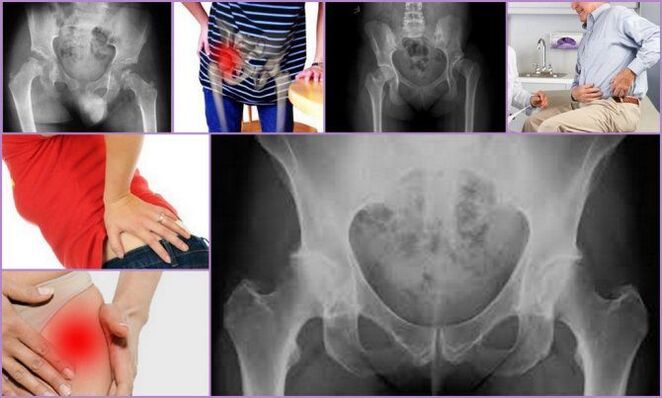
Diagnose
If signs of coxarthrosis occur, you should contact a chiropractor as soon as possible. Initially, the doctor will interview the patient and find out the nature of the complaints, then will conduct an examination and perform functional tests, comparing the length of the leg. As a rule, the data obtained are enough to tell with a high degree of confidence the presence of coxarthrosis.
But since such a clinical picture can be accompanied by a number of other diseases of the hip, both inflammatory and non-inflammatory in nature, instrumental diagnostic methods are required. With their help, a specialist will be able not only to determine the presence of coxarthrosis, to distinguish it from lens syndrome caused by spinal pathology, but also to accurately assess the extent of coxarthrosis. its development, which means choosing the most effective treatment strategy.
Today for the diagnosis of coxarthrosis are used:
- X-ray of the hip - the resulting image allows you to detect signs of destructive changes, the presence of osteoclasts, the nature of the deformation of the bone structure and measure the thickness of the joint space.
- CT is a more modern method for diagnosing bone diseases, providing clearer data than x-rays, but is more expensive. Therefore, CT is indicated in controversial cases, when it is necessary to clarify the diagnosis and extent of hip destruction.
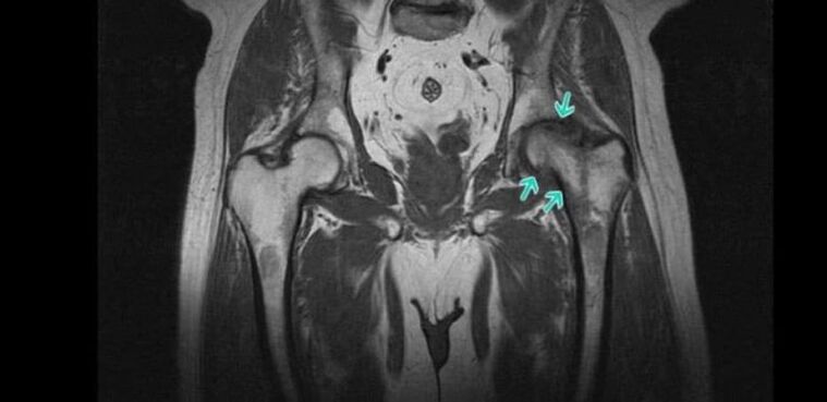
- MRI is a highly informative method of examining joints, providing the maximum amount of information about the state of the joint and all its structures, especially the hyaline cartilage, ligaments and arch features. blood supply.
Patients are assigned a number of laboratory tests, including KLA, OAM, rheumatic tests, biochemical blood tests, and others.
Conservative treatment of coxarthrosis

When coxarthrosis grade 1 or grade 2 is diagnosed, treatment is carried out conservatively. For each patient, they were selected individually, taking into account the detected comorbidities. Therefore, it may be necessary to consult not only an orthopedist, but also a doctor of other specialties, who will select the necessary treatment to combat concomitant diseases.
As part of the treatment of coxarthrosis, the patient is prescribed:
- drug treatment;
- exercise therapy;
- physical therapy.
It is imperative that all patients take measures to eliminate the effects of factors that increase the load on the leg and contribute to the progression of degenerative changes in the hip. This includes adjusting your diet and increasing your level of physical activity if you are overweight. If the patient frequently experiences excessive exertion, the type of activity should be changed or the intensity of exercise reduced, if the load is due to sports. In some cases, special bandages and orthotics should be used to immobilize the hip and remove it during exertion.
Medical therapy
As part of drug therapy, the patient is selected for separate drugs, taking into account existing concomitant diseases. As a rule, drugs of the following pharmacological groups are indicated for coxarthrosis:
- NSAIDs - a wide group of drugs with analgesic and anti-inflammatory effects (there are many dosage forms, including tablets, capsules, gels, creams, injection solutions, allowing you to choose an effective form of application. most efficient and convenient);
- corticosteroids - drugs with strong anti-inflammatory effects, but due to the high risk of side effects, especially when used in the oral form, should only be prescribed for short-term courses in the form of injections;
- muscle relaxants - drugs that help reduce muscle tone, allowing you to effectively cope with muscle spasms, which are often seen in coxarthrosis;
- chondroprotectors - a group of drugs that contain components used by the body to regenerate cartilage tissue;
- preparations that improve microcirculation - help improve the nutrition of soft tissues and activate metabolic processes in the affected area;
- Vitamins of group B - are indicated for disorders of nerve conduction due to nerve compression by altered components of the hip joint.
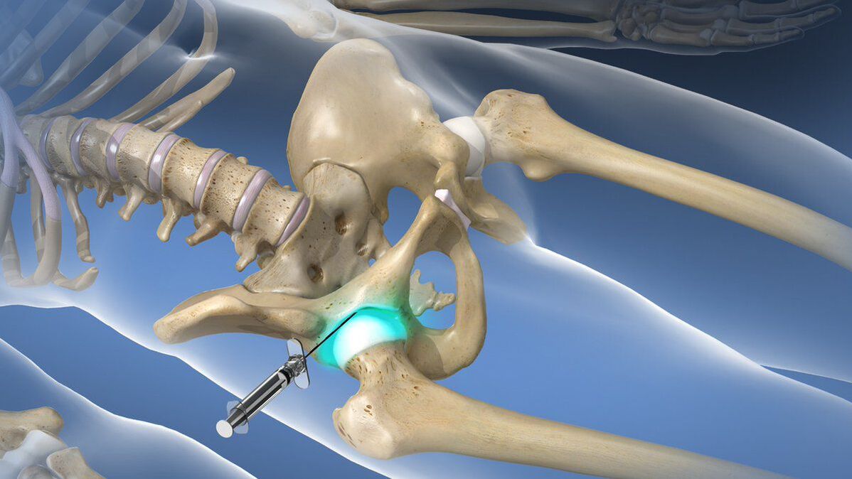
If coxarthrosis causes acute pain that cannot be stopped with the help of a prescribed NSAID, intra-articular or periarticular blockade should be administered to the patient. Its essence lies in the direct injection into the hip cavity of an anesthetic solution in combination with corticosteroids. This will allow you to quickly eliminate pain and reduce the inflammatory process. But the blockade can only be carried out by a qualified medical staff in a specially prepared room. Performing such procedures at home is not shown.
exercise therapy
When coxarthrosis is diagnosed, regular exercise therapy is imperative. Similar to drug therapy, the set of therapeutic exercises for each patient is selected individually, taking into account the degree of destruction of the hip, the degree of physical development of the patient, the nature of the diseases. concurrently (particular attention is paid for cardiovascular pathologies).
Thanks to daily exercise therapy, you can:
- reduce the severity of pain;
- increased mobility of the hip joint;
- reduce the risk of muscle atrophy;
- eliminate the spasm of the thigh muscles;
- activates blood circulation and thus improves the nutrition of the affected joint.
All exercises should be performed in a rhythmic manner, avoiding sudden and jerky movements. But if pain appears during exercise therapy, you should definitely contact your doctor to correct the selected complex or conduct a re-diagnosis to rule out disease progression and the need forsurgery.
Physical therapy
Comprehensive treatment of coxarthrosis includes physical therapy courses that have anti-inflammatory, analgesic, decongestant, and tonic effects on the body. Therefore, most often patients are prescribed 10-15 procedures:
- therapeutic ultrasound;
- electrophoresis;
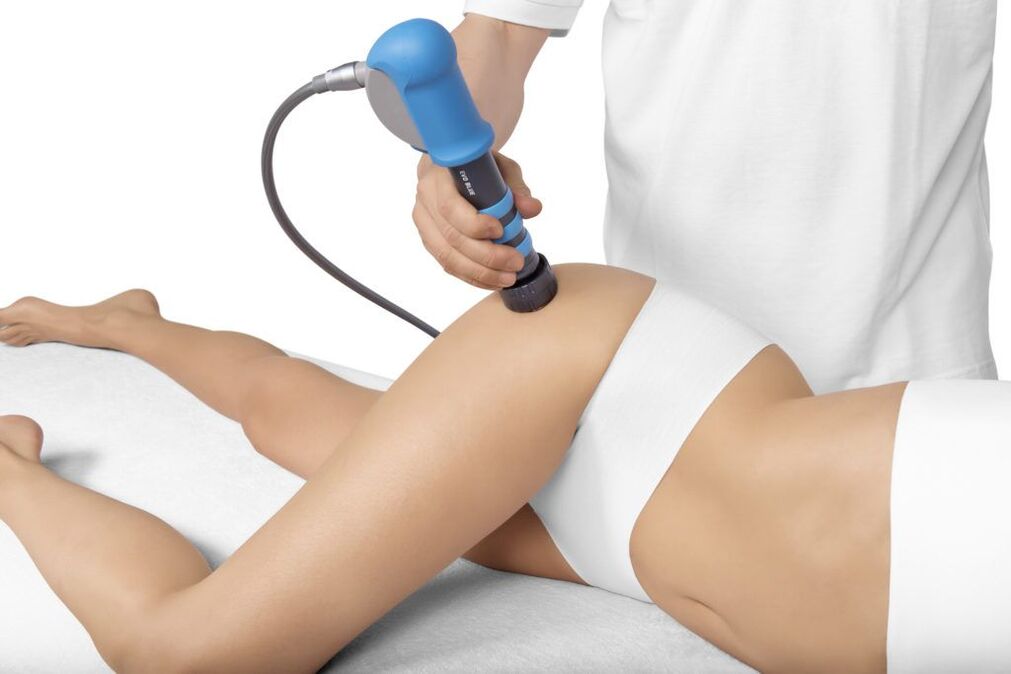
- UVT;
- acupuncture therapy;
- laser therapy, etc. v.
Recently, plasmolifting has been increasingly used as part of the conservative treatment of coxarthrosis, which can significantly increase the rate of hyalinized cartilage regeneration. The essence of the procedure is the introduction of purified plasma into the hip cavity, which is obtained by centrifugation from the patient's own blood.
Coxarthrosis surgery
If a patient is diagnosed with grade 3 coxarthrosis, he will be indicated for surgical intervention, since conservative methods in these cases have been impotent. Unfortunately, such situations are extremely common today, as a large number of patients seek medical help when they can no longer tolerate the pain or have severe mobility limitations that cause them to lose weight. ability to work and move independently.
Timely surgical intervention can completely eliminate these disorders and restore the patient's normal mobility, significantly improving the quality of life. The signs to do it are:
- significantly reduce joint space by more than 80%;
- the presence of severe pain in the hip joint, which cannot be eliminated;
- pronounced movement disorders;
- femoral neck fracture.
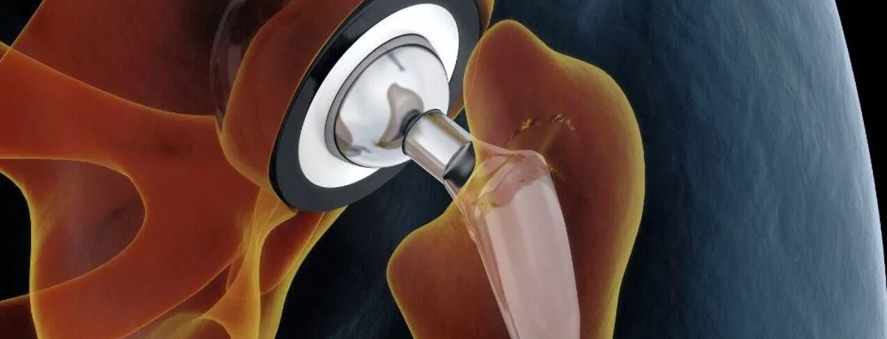
The gold standard for the treatment of severe coxarthrosis, including in the elderly, is hip arthroplasty. This surgery involves replacing the destroyed hip joint with an artificial organ made of durable and biocompatible materials at the same time. Internal medicine allows you to fully restore the function of the hip joint, eliminate pain and return a person to a full-fledged active life.
The essence of this type of surgical intervention is the removal of the femoral head and a small piece of the neck. In addition, the surgeon will need to prepare the surface of the pad for endocytosis, i. e. remove all the bone cells that have formed and achieve maximum restoration to its normal shape. . After that, an endocardium of the selected type is installed, which is fixed with special cement (preferably for the treatment of the elderly) or by non-cement. In the second case, the intraosseous bone has a special spongy part in contact with the bony structures. Its fixation in the acetabulum is provided by the germination of bone tissue through the sponge.
For each patient, the joint type is selected individually. The most effective is total arthroplasty, which involves the complete replacement of the entire hip, i. e. the neck and head of the femur, as well as the rotator cuff.
If the patient has preserved normal hyaline cartilage on the surface of the disc, the patient can undergo partial arthroplasty with only femoral head and/or neck replacement. For this purpose, endoprosthes of different designs are used: unipolar and bipolar.
The only disadvantage of arthroplasty can be considered as the need to replace the installed endoscope after 15–30 years.
After endothelial replacement, the patient is restored to function, the recovery time depends on the rate of tissue repair. As part of the recovery process, exercise therapy, physical therapy, and therapeutic massage are prescribed.
Before the advent of modern endoprosthes, patients with grade 3 coxarthrosis were indicated for osteotomy or arthroplasty. These techniques are used less and less these days, as they have several disadvantages. So, hip joint involves fixing the bony structures of the hip joint with metal plates. As a result, the pain syndrome is completely eliminated but the joint loses its mobility completely. Therefore, after joint treatment, the patient can only stand but is no longer able to walk independently because the hip joint cannot be moved. So today arthrodesis is practically not performed.
Osteotomy involves performing an artificial fracture of the femur with a combination of bone fragments such that the load on the affected hip is relieved. But the surgery only gives a short-term effect, in the future the need for orthopedic surgery still arises.
Thus, degenerative hip disease is a rather dangerous disease, which can cause disability. It severely reduces the quality of life and makes a person unable to work. But if you pay attention to the early signs of pathology and get advice from a chiropractor in time, you can slow down its progression and significantly improve your state of health. But with active coxarthrosis, there can only be one solution - arthroplasty. Fortunately, this method can be used even with severe degenerative-dystrophic changes and completely restores normal function of the hip.